Snails in 3D
Understanding what organs are present in animals and how they differ between related species is crucial for understanding the biology and evolution of different species.
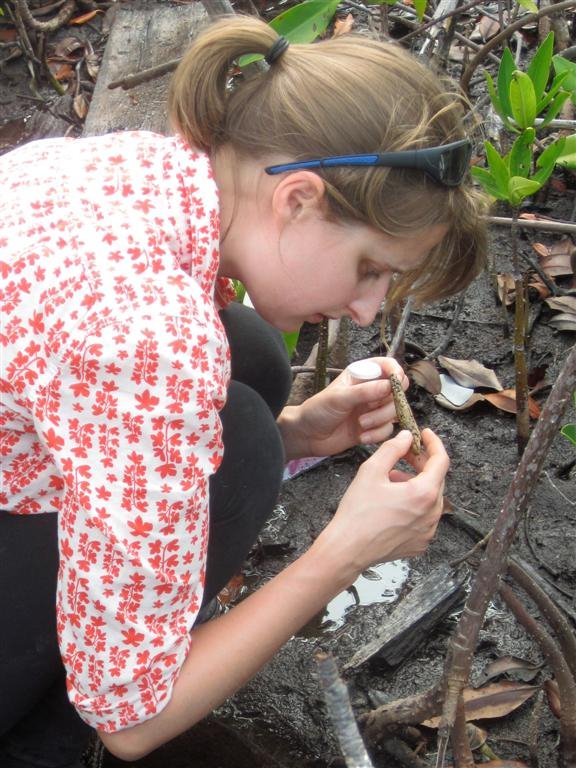
Dr Rosemary Golding in the field.
Image: Tim Rawlings© Tim Rawlings
Traditionally, scientists use micro-dissection, using ultra-fine surgical instruments and a microscope, to explore the internal anatomy of snails and other small invertebrate animals. But some Australian snails are just too small to be cut open and inspected, and one species in particular, at just one millimetre long, caught my attention as I examined the Museum’s collection of specimens.
Based on its external shell, it seemed to be a new species found in intertidal algae around Sydney. I really needed some information on its internal organs in order to understand what it had in common with other Australian species.
To solve this problem, I turned to a new technique that can build a three-dimensional (3D) model of the internal anatomy of tiny specimens. This procedure, known simply as 3D reconstruction, was developed by other scientists, particularly a group of German researchers led by Dr Bernard Ruthensteiner at the Bavarian State Collection of Zoology, who wanted to know more about the microanatomy of some fascinating but tiny snails.

© Australian Museum
3D reconstruction
The process begins by taking very fine slices of one animal embedded in plastic. Fortunately for my work, these histological sections had already been created by a Swedish researcher, Dr Anders Warén, who had visited the Australian Museum in the 1980s.
Using a high-resolution camera connected to a microscope, I took digital photographs of these sections and stacked the images one on top of the other in the correct sequence to create a computerised ‘virtual model’ of the animal.
After carefully tracing the outlines of the organs I wanted to see in 3D on each image, I employed special software to build models that could be rotated and explored on screen. These models showed the organs in much greater detail than could be revealed through dissection alone. They also helped me show that this new species is unique in Australia, and is in fact more closely related to a group of deep-sea snails found in Antarctica and Norway!
The technique of 3D reconstruction is an exciting breakthrough in invertebrate anatomy, which can be an incredibly useful tool for understanding biodiversity, evolution and even basic biology.
Story by Dr Rosemary Golding, Chadwick Biodiversity Fellow
(The full version of this article appeared in a recent issue of the Australian Museum's Explore magazine.)
